The gills of Coenobita and Birgus are modified for air-breathing but are reduced in number and size and have a comparatively small surface area. The branchiostegal lungs of Coenobita (which live in gastropod shells) are very small but are well vascularized and have a thin blood/gas barrier. Coenobita has developed a third respiratory organ, the abdominal lung, that is formed from highly vascularized patches of very thin and intensely-folded dorsal integument. Oxygenated blood from this respiratory surface is returned to the pericardial sinus via the gills (in parallel to the branchiostegal circulation). Birgus, which does not inhabit a gastropod shell, has developed a highly complex branchiostegal lung that is expanded laterally and evaginated to increase surface area. The blood/gas diffusion distance is short and oxygenated blood is returned directly to the pericardium via pulmonary veins. We conclude that the presence of a protective mollusc shell in the terrestrial hermit crabs has favoured the evolution of an abdominal lung and in its absence a branchiostegal lung has been developed. [1]


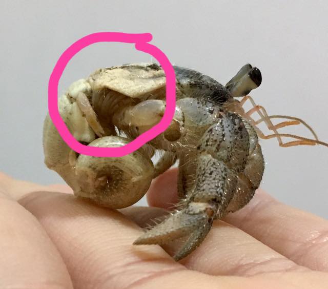
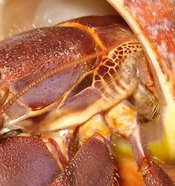
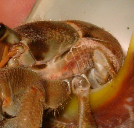
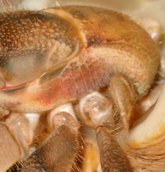
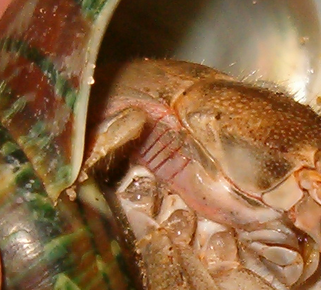
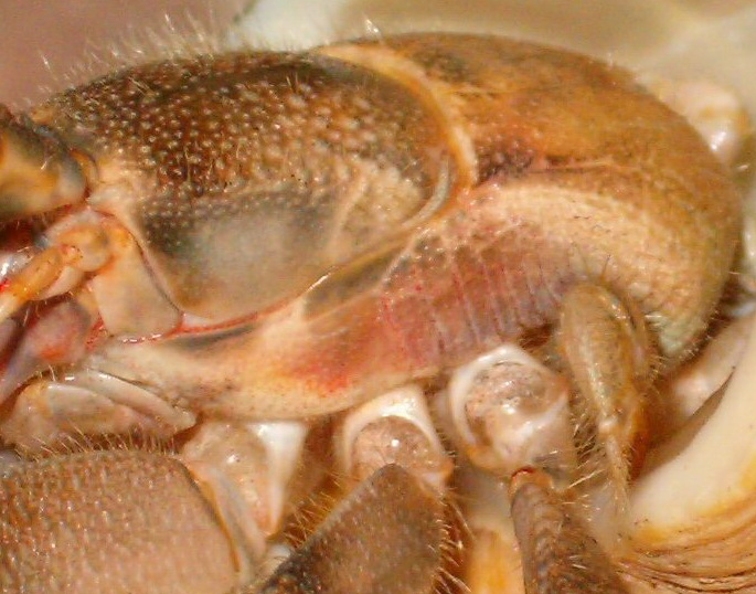
We are building image galleries of specific body parts. If you have high resolution, clear photos that you would like to donate to this project please upload your photos to Dropbox here: https://www.dropbox.com/request/ajUgzmoG7co86iH2X0fh
Overview of the anatomy of a land hermit crab (Coenobita)
References:
1 The morphology and vasculature of the respiratory organs of terrestrial hermit crabs (Coenobita and Birgus): gills, branchiostegal lungs and abdominal lungs
C.A. Farrellya, P. Greenawayb, ,
a 32 Tramway Pde. Beaumaris, Vic. 3193, Australia
b School of Biological, Earth and Environmental Sciences, University of New South Wales, Sydney, N.S.W. 2052, Australia

